Case discussion, teaching and specialty training on the same section.
A cloud application for digital pathology, with an interface that follows the workflow of the department and needs no lengthy training.
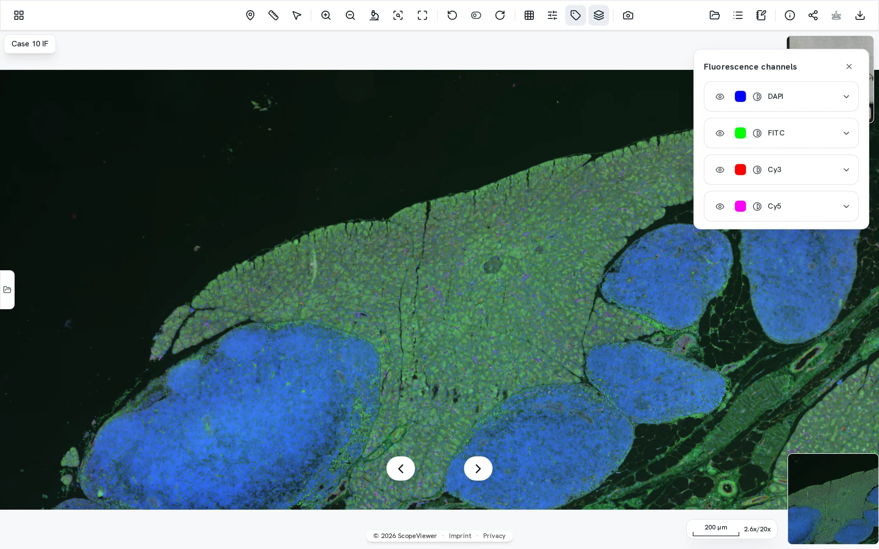
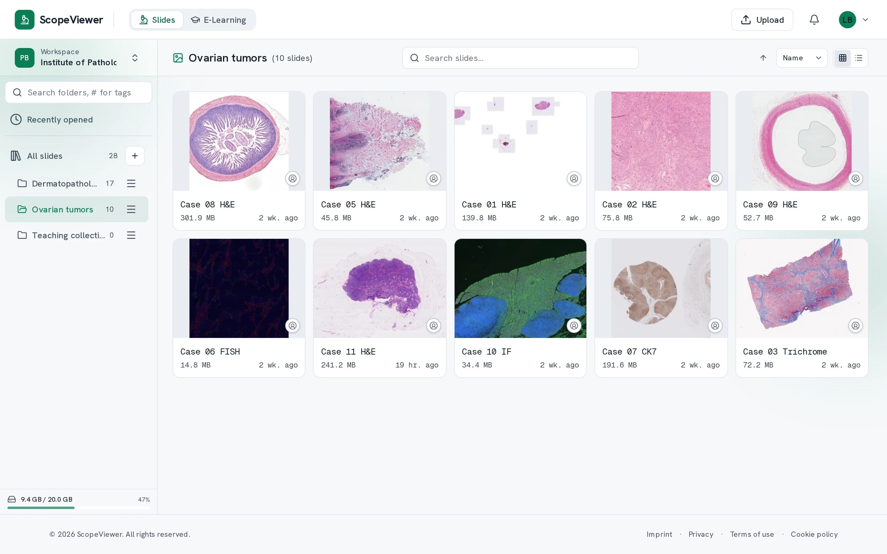
View multichannel fluorescence sections channel by channel or all overlaid, each channel with its own colour, brightness and contrast.
One slide collection for everyone at your institution.
Filing, sharing and discussing a case all happen in the same place.
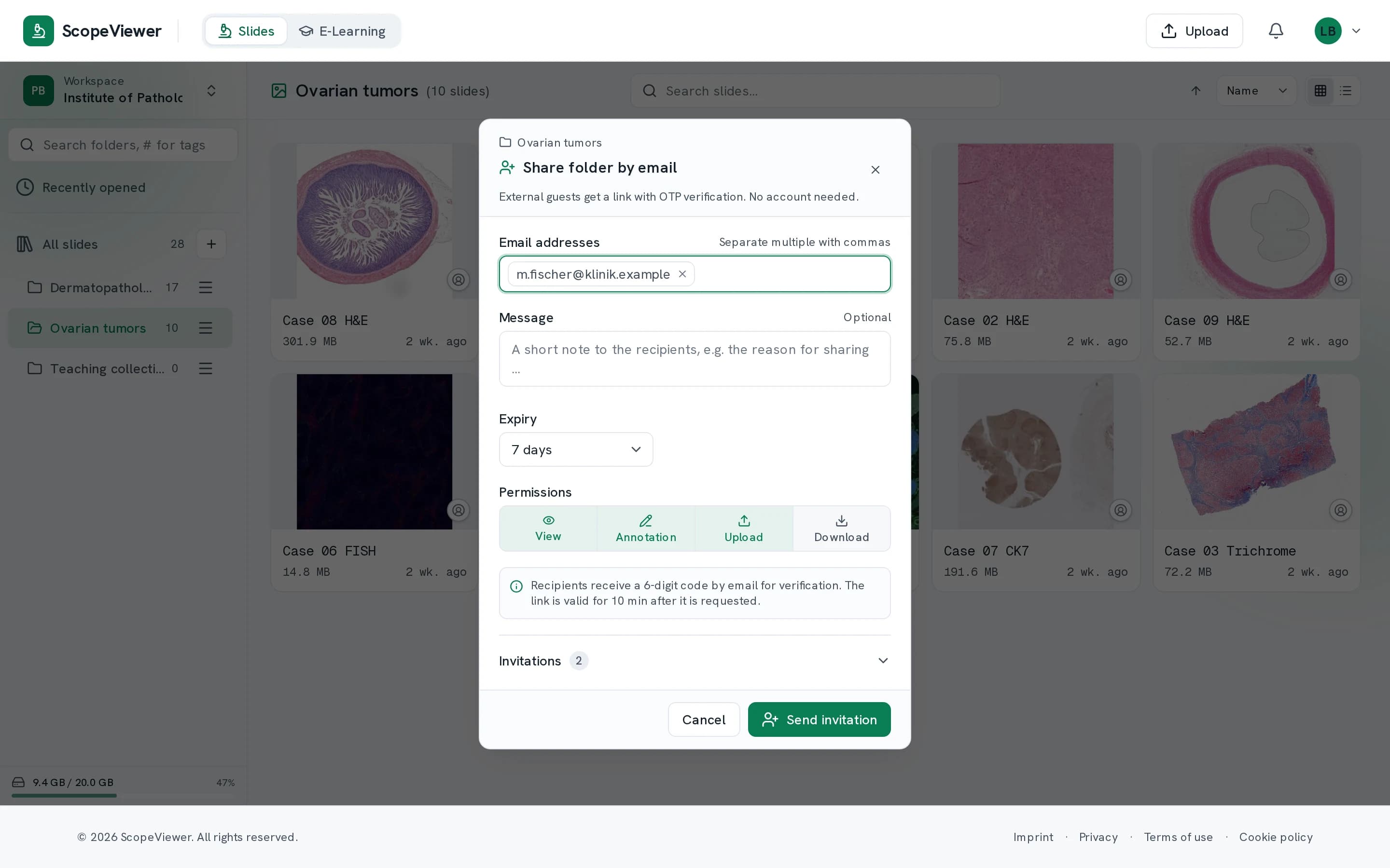
- You share outwards by link. Nobody needs an account for it, and you can withdraw the share at any time.
- Markers, measurements and notes are there for everyone else the moment you make them.
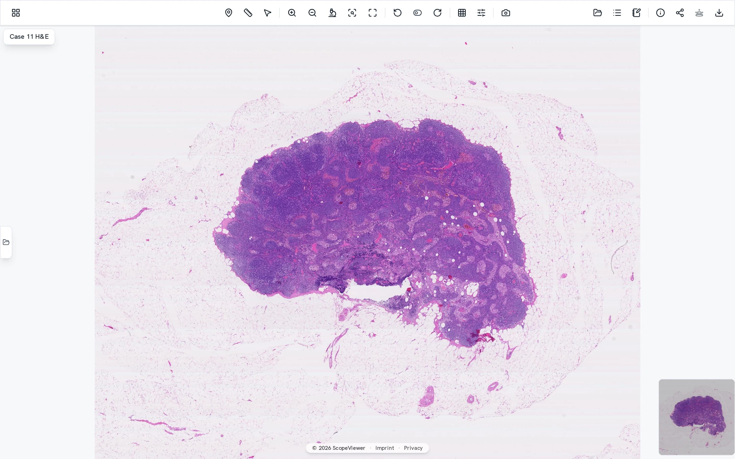
- Up to nine views stay coupled, keeping H&E and immunohistochemistry in register.
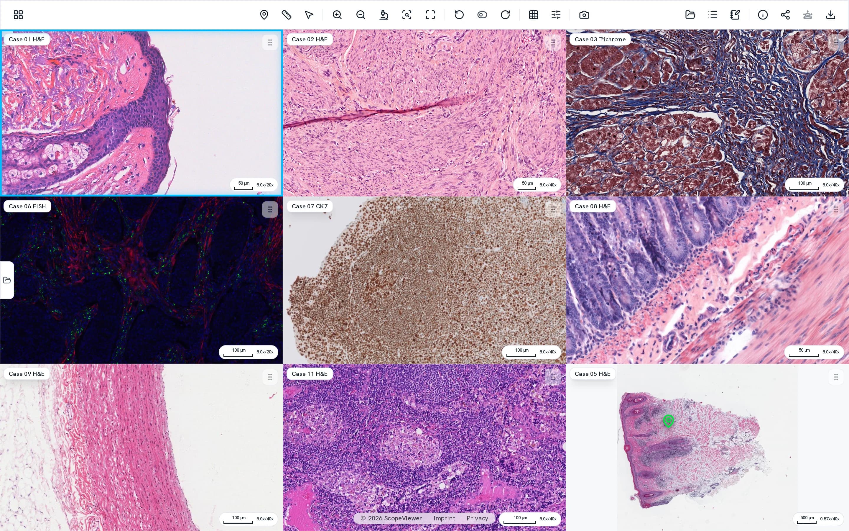
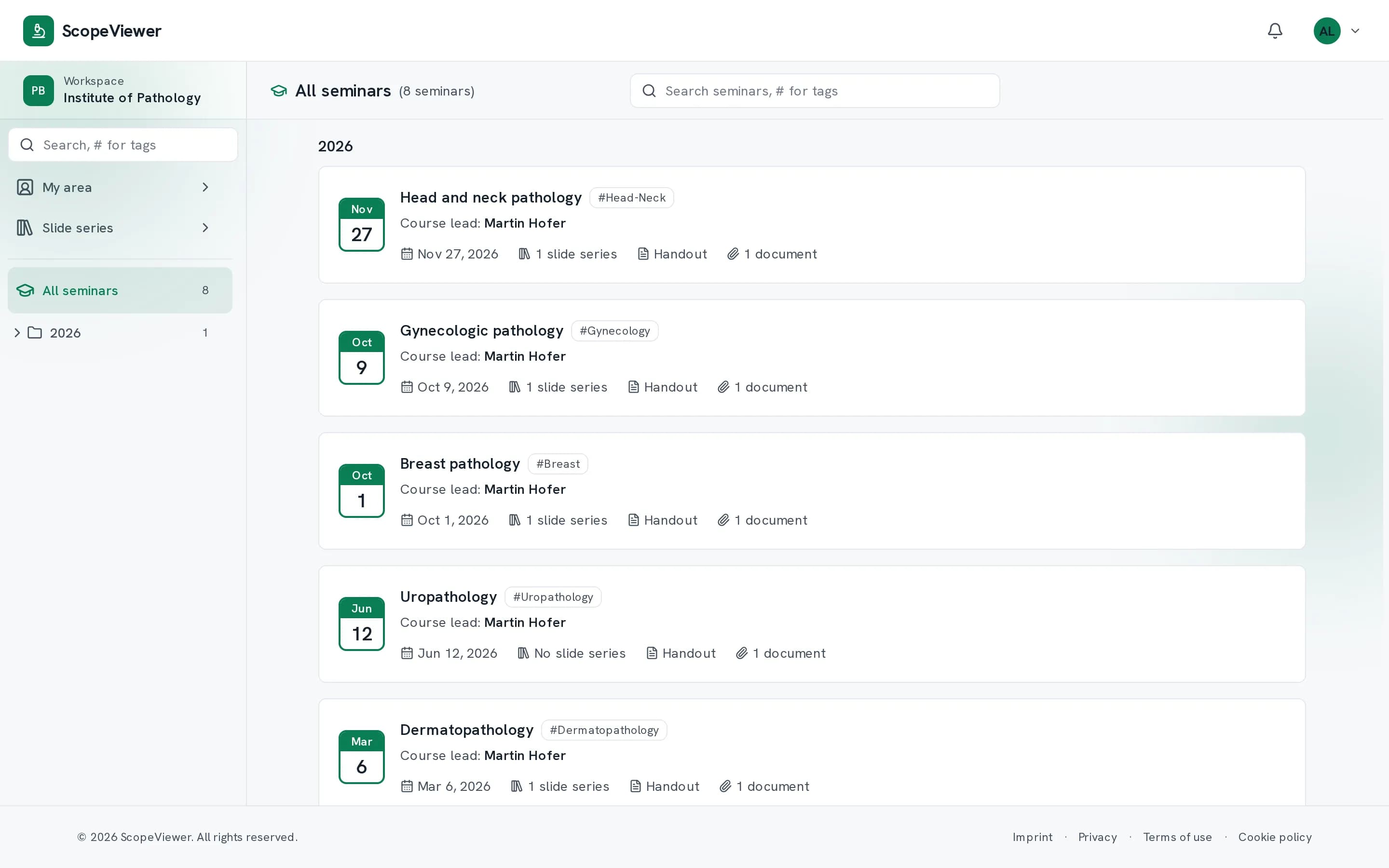
Teaching needs no second platform.
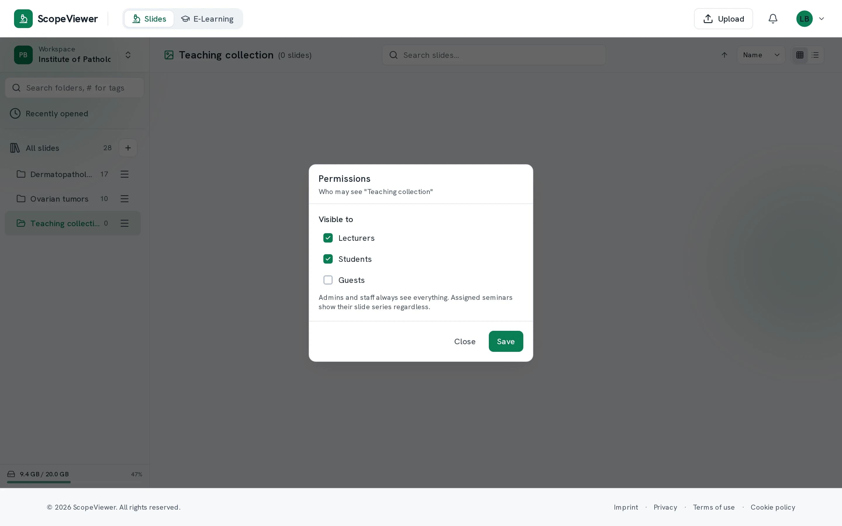
What you put in a folder is what participants see in the course, released on the date you set.
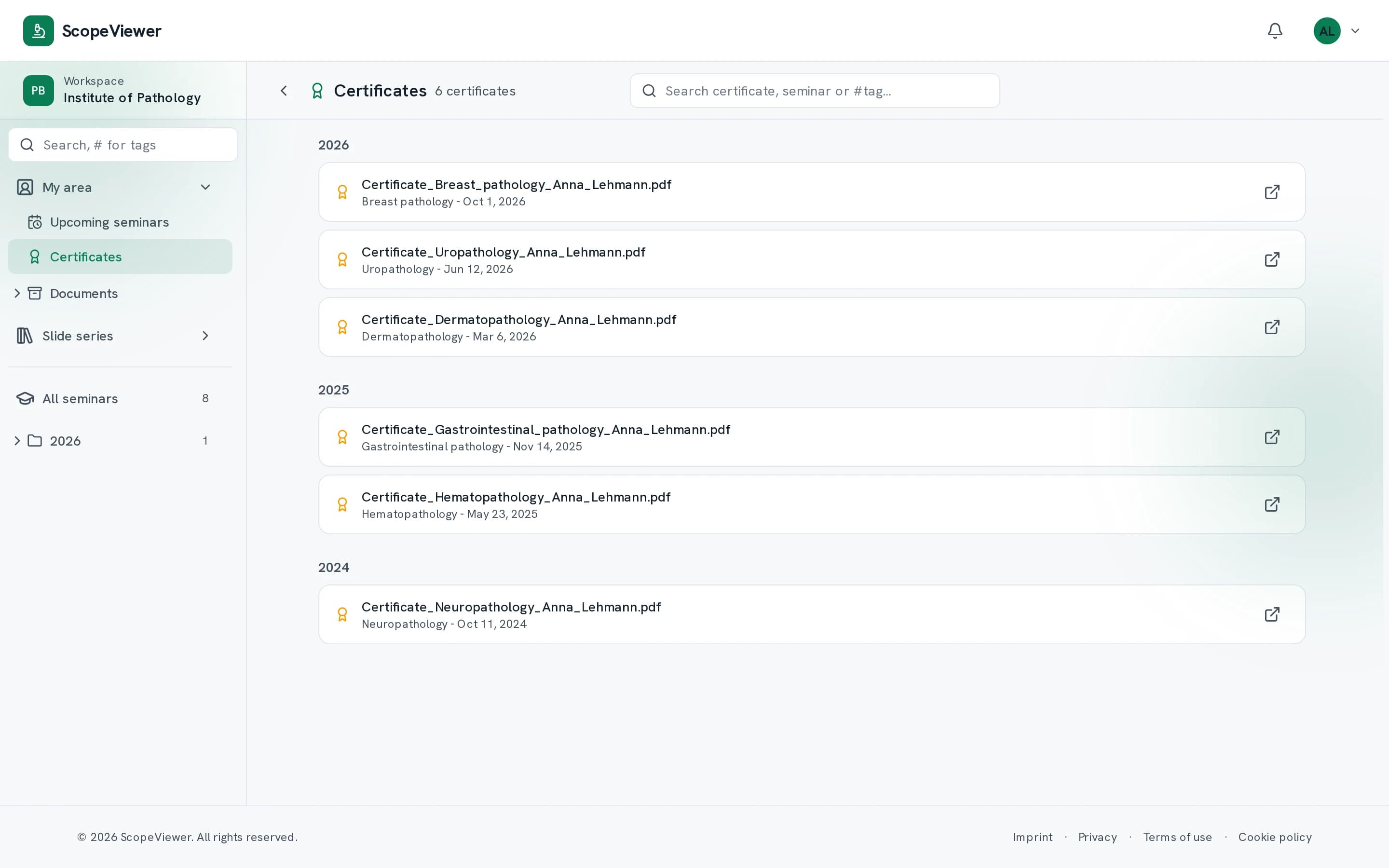
Each person gives their own diagnosis for each case, up to the deadline shown in the course. Attendance certificates go in as a single collected PDF, each one matched to its recipient for you.
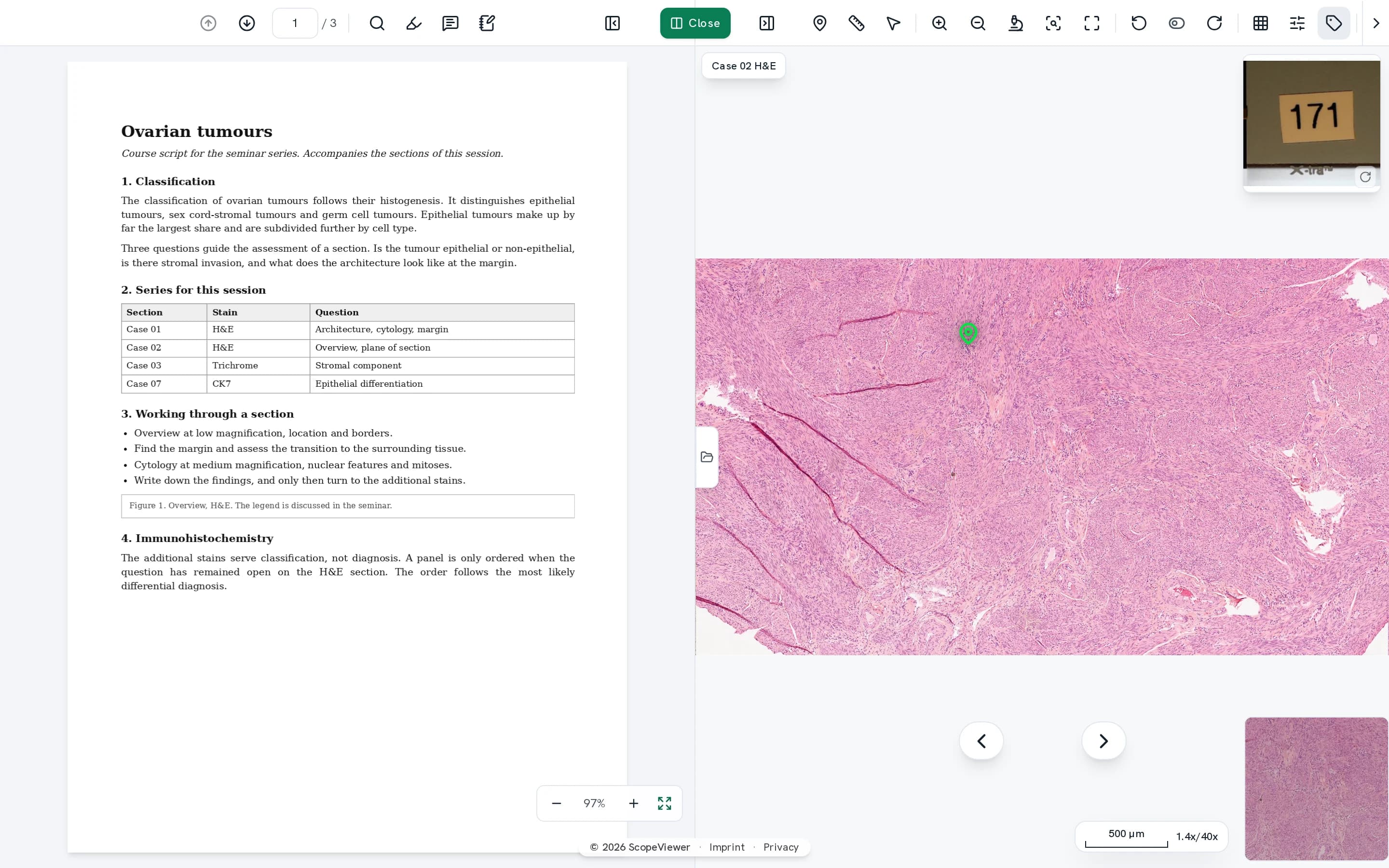

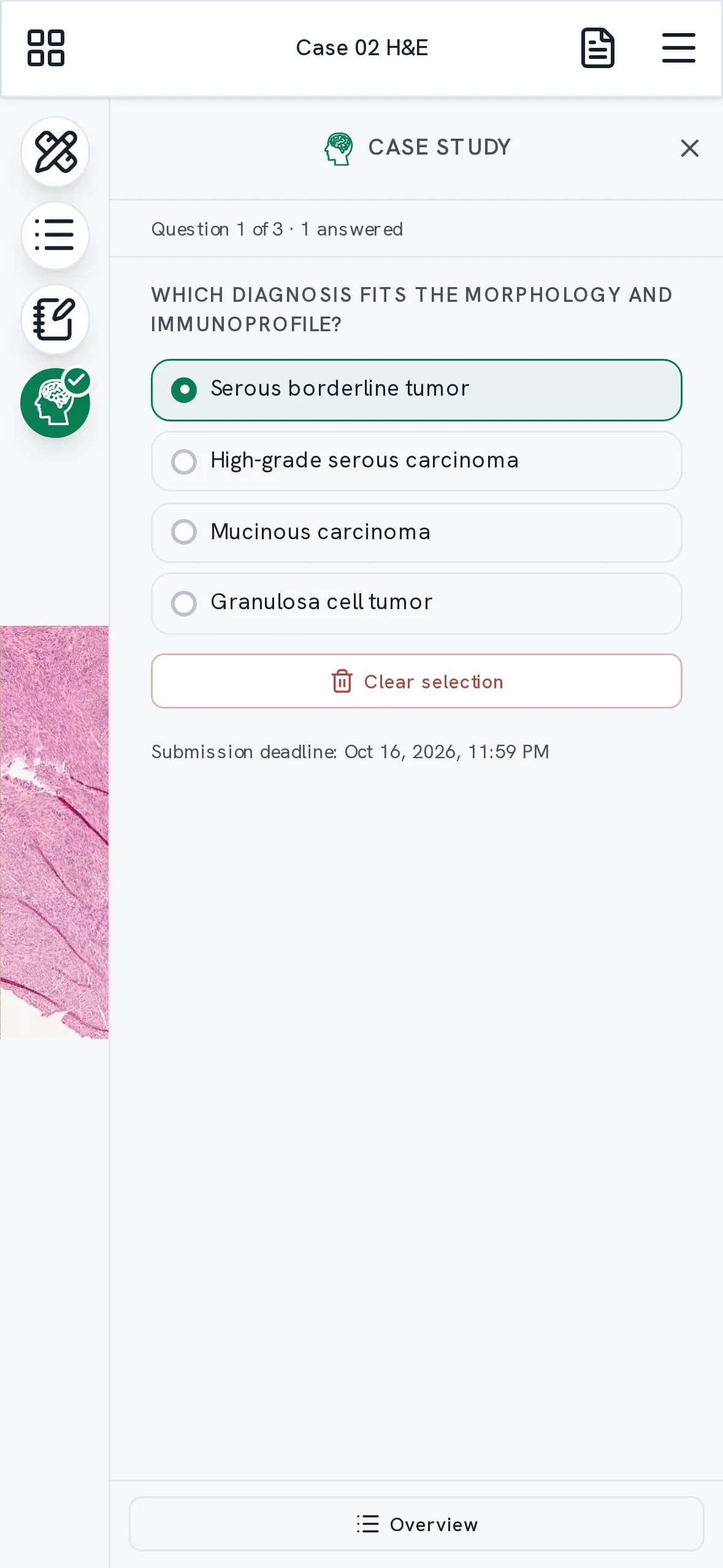
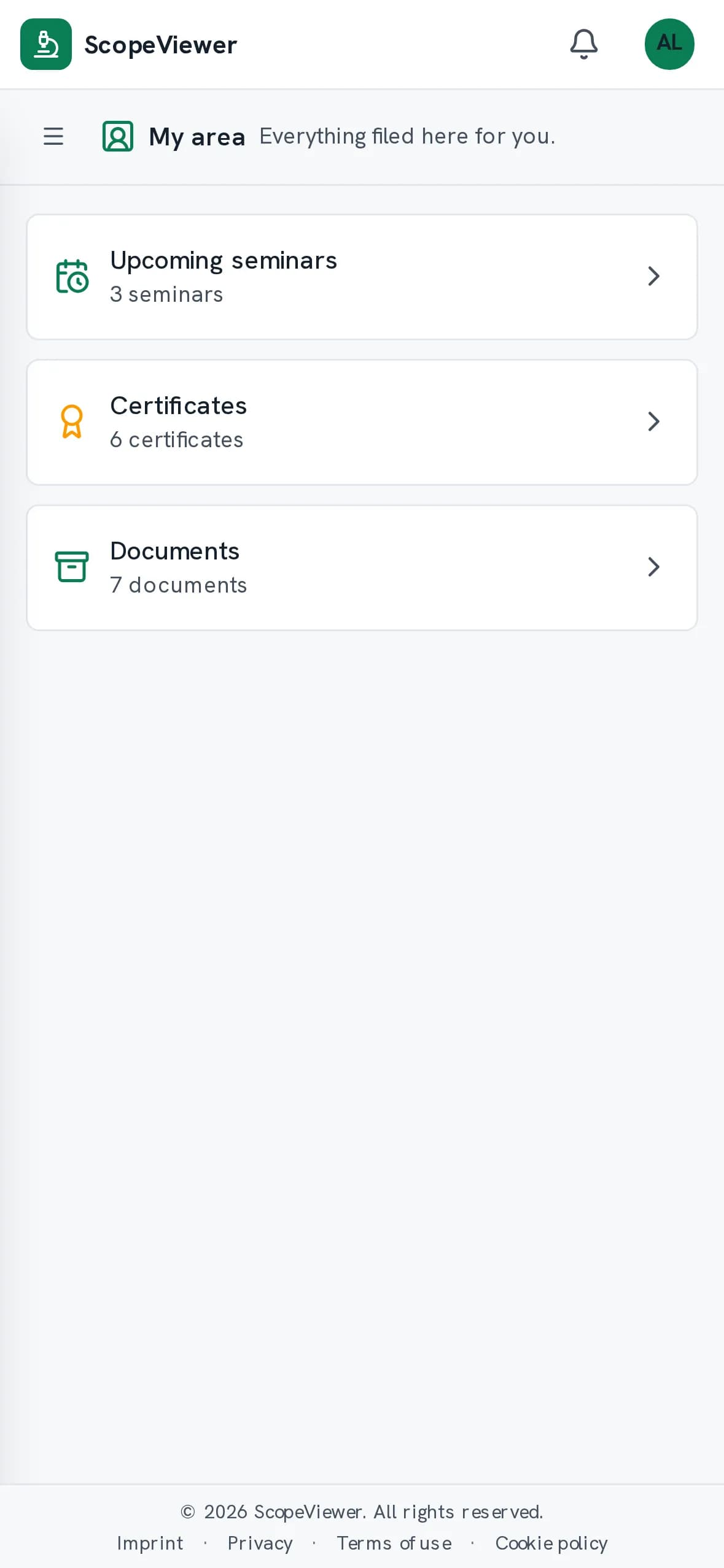
Away from the desk, you carry on.
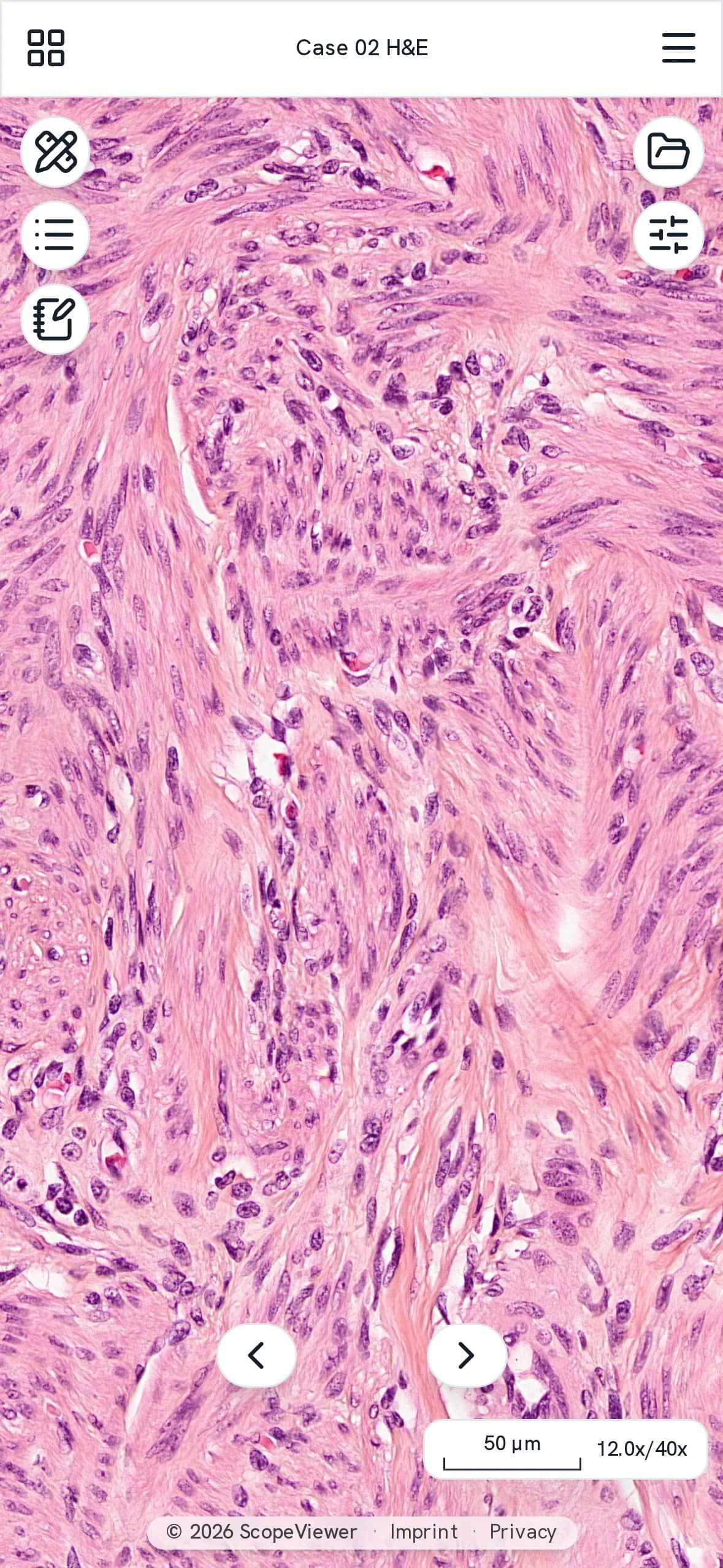
Whatever catches your eye, you can mark on the phone there and then. The section is there at full resolution.
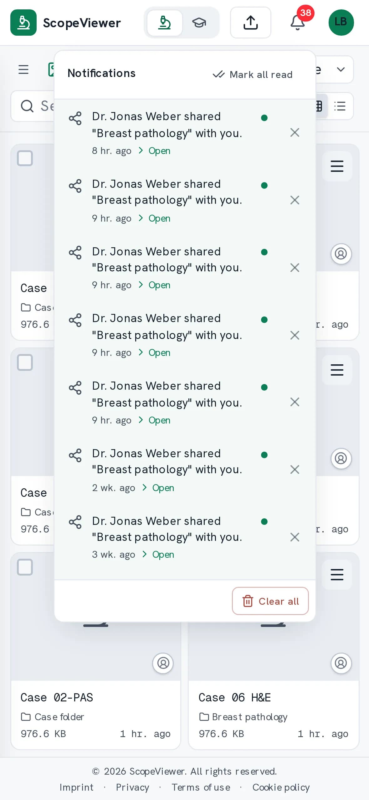
Your training carries on here too, right through to a submitted diagnosis. When a course is assigned or a document filed for you, a push notification reaches your phone and your computer, once you allow it.
Nine things that come up before the decision.
- The archive stays findable
- Folders carry keywords such as organ, stain or study, from one list that applies across the institution. Rename one and every folder follows, and someone who did not file the case can still find it again.
- Supported formats
- Aperio, Hamamatsu, 3DHistech, Leica, Olympus, Philips, Ventana and DICOM open just as the scanner saved them, even when one section is a whole folder of files. We add further formats whenever you need them.
- Videos for your course only
- We host your lectures and teaching videos, protected against downloading. Apart from your team, only the people you assign to the course can play them. You don't need a contract with a video service of your own.
- Sharing that runs both ways
- Colleagues outside your institution put their own sections into the shared folder instead of sending them to you. They can only remove what they brought in, and their sections then take the same route as your own.
- Fluorescence in the same viewer
- Fluorescence slides sit in the same folder as your brightfield slides and open in the same viewer. Where several focal planes were scanned, you see every FISH signal in one image without focusing through them.
- Several sites under one account
- Working at two institutions takes a single account. Each site keeps its own roles, its own storage and its own courses, and what sits in one stays invisible in the other. You switch inside the account.
- A whole team at once
- You set up a whole team from a CSV file, up to 500 people at once, with one role for the whole import. The invitations go out by email. Anyone who signs up later on their own waits for your approval to join.
- The handout is worked in
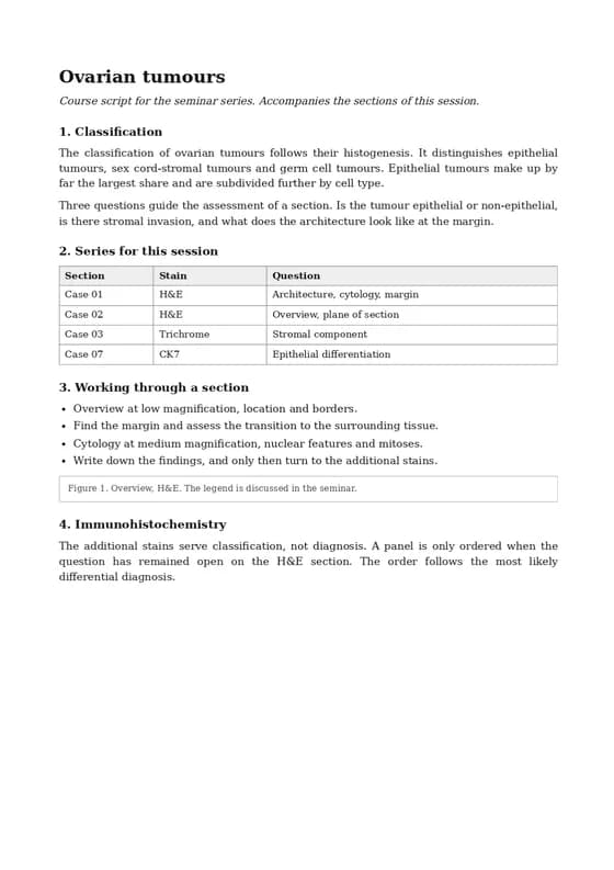
- However long the handout, it is searched as participants type, and they read it beside the section. What someone highlights and notes in it is seen by no one else, not even by the person leading the course.
- The same section only once
- Uploads are compared by the file's checksum, not by its name. If the section already exists in your institution, you see its name and its folder and jump straight there instead of storing it a second time.
What your IT department wants to know before it signs off.
- Servers and data stay in the EU, processed under the GDPR.
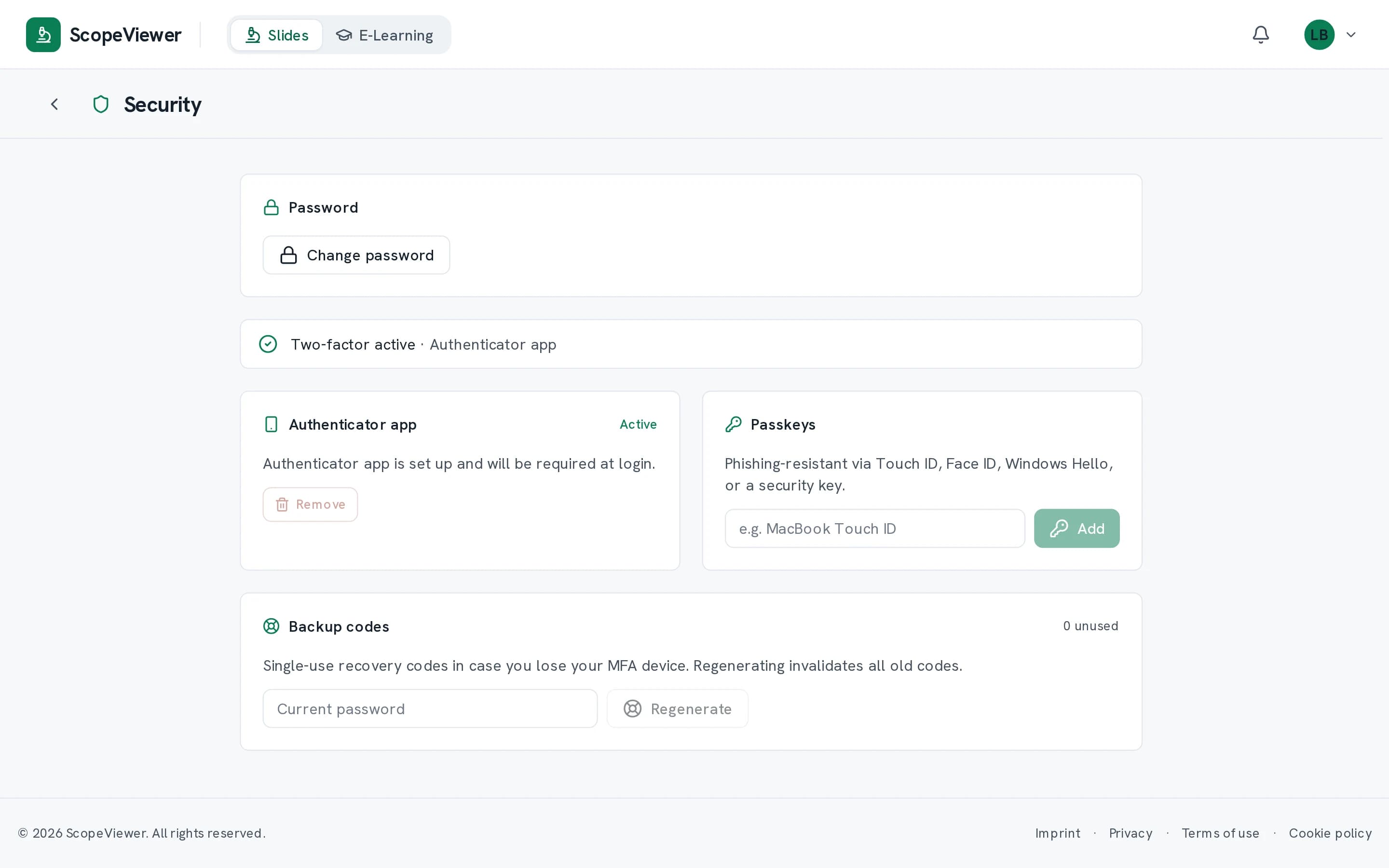
- Admin accounts always need a second factor, and you can require it of everyone.
- One session stays active per account, so a shared password shows up at once. Unattended workstations sign out after five minutes to eight hours.
- Remove an account and access ends at once, not at the next sign-in. What they created stays with your institution.
- A forwarded invitation is of no use to anyone, and it expires after thirty days.
- Sign-ins and changes sit in a trail of your own, including every visit from our team. Entries are only ever added, and nobody can delete them.
Request access
ScopeViewer is provided by invitation. Send us a short message and we will set up access for your institution.